Developmental Biology
/Parastichopus parvimensis
Auricularia


Auricularia
Skeleton
Tripartite Gut
Coelom Formation
Haline Bodies
In this larval stage, first, the tripartite gut fully forms. The archenteron elongates - the top forms the mouth, the middle the esophagus, and the bottom the stomach and rectum (Figure 1)(Figure 2). Later, the top of the archenteron opens up at the mouth, and the rectum curves around to face the same direction as the mouth.
Another important feature of developing larvae is the ciliary band, one continuous band looped in a complicated pattern around the edges of the larvae (Figure 5). The ciliary band, as its name suggests, is composed of cilia, which the larva uses to propel itself through the water. All cilia beat away from the oral hood (Lacalli & West, 2005). For more information on auricularia physiology of the ciliary band and tripartite gut, see Compound Microscopy.
The formation of the tripartite gut transitions the gastrula into its first larval stage, called an auricularia. Instead of the animal vegetal axis, auricularia are oriented around two new axes, the dorsal ventral axis and anterior posterior axis The dorsal ventral axis can be considered the “front back” axis, with the openings of the mouth and anus facing towards the ventral side (Figure 3). The animal vegetal axis becomes the anterior posterior axis respectively (Figure 4).
Like early development, all stages of auricularia development occur in the water column. Auricularia are planktotrophic larvae, meaning that they feed in the water column (versus lecithotrophic larvae). They are also indirect developers (versus direct), meaning that they have larval forms much unlike their adult forms, that metamorphose. As a result of indirect development and planktotrophy, the larva spend more time in the water column than their lecithotrophic counterparts would. Although it is unknown as to how long Parastichopus parvimensis auricularia float in the water column in the wild, previous studies observed auricularia stages lasting from anywhere between 17-35 days, or 2 to 4 months before metamorphosis to doliolaria stages, depending on culturing techniques and diets (Cameron, 1980)(Smiley 1985)(Lacalli & West 2000). In our study, auricularia stages lasted for as short as 16 days before metamorphosis. They were fed a combination diet of green phytoplankton, Dunaliella tertiolecta, and red phytoplankton, Rhodomonas lens.
A distinct feature of echinoderm development is coelom formation. Classic coelom development begins with an axocoel, hydrocoel, and somatocoel, located in descending order around the tripartite gut. Holothuroids are thought to lack axocoels. Recent studies, however, suggest that holothuroids do indeed possess an axocoel, fused with the hydrocoel to form the axohydrocoel (Balser et al. 1993)
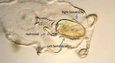
In late auricularia, holothuroids have three coelom compartments: a hydrocoel and paired somatocoels. The hydrocoel is the most conspicuous coelom, located on the left side of the larva next to the lower esophagus. The somatocoels are much smaller, and are located on either side of the stomach. The left somatocoel is usually a bit larger than the right somatocoel (Figure 6). Occassionally, the left and right symmetry is switched with the hydrocoel and larger somatocoel on the right instead of the left. In addition, the left hydrocoel opens towards the dorsal side of the larva, through the coelomoduct, called the hydropore (Figure 7).
In late auricularia phases, small spheres called haline bodies (also known as hyaline spheres) appear. Haline bodies are unique to holothuroid development, although little is known about their function or development. Late auricularia contain up to 10 haline bodies – 5 symmetrically placed on each the left and right sides of the larva. Occasionally, fewer than 10 haline bodies occur, which has been correlated to levels of feeding and diet (Morgan 2008).
There is much to be studied about haline bodies, as both the function and order in which they are appear go relatively unstudied. In this study, haline bodies were first spotted in locations (1) and (2) quite consistently (Figure 8).
Lastly, a developing skeletal structure can be observed in late auricularia. This skeletal structure is the beginnings of the adult skeleton (Figure 9).
Figure 1: Early tripartite gut - (a) mouth
(b) esophagus (c) stomach and rectum
Figure 2: Slightly more developed tripartite gut
Figure 4: Anterior Posterior Axis
Figure 3: Dorsal Ventral Axis
Figure 5: Auricularia
Figure 7: Hydropore from Left Coelom
Figure 6: Coeloms
Figure 8: Haline Bodies
Hydrocoel: wraps around the esophagus and forms the ring canal and radial canals of the water vascular system
Hydropore: forms the madreporite of the water vascular system.
Left and Right Somatocoels: form the perivisceral coeloms
Coelomic Fates:
Figure 9: Skeletal Structure at Posterior end